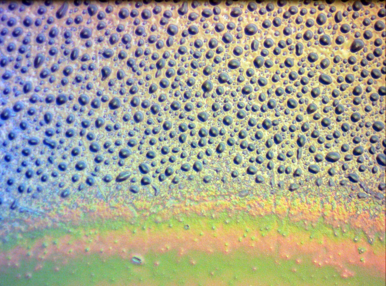
Differential interference contrast is a polarization based technique that induces lateral shear by less than the resolution of the system, interfering the two beams which causes edges, primarily, to become much more visible than a brightfield image. The contrast enhancement does not suffer from the halos present with phase contrast. The above image is of a diffusing filter glass, in reflected light DIC with 20x M Plan.

The diagram above is from Nikon's Microscopy U
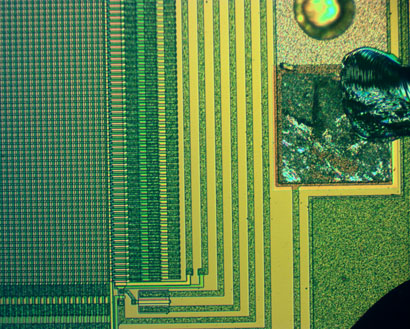
Here is an animation of rotating the analyzer (9.8 MB) by about 10 degrees per frame, with 18 frames, of a bare CCD chip at 10x with M Plan in reflected light DIC. The left side shows the actual pixels, which are 15 microns square (3 color CCD).
While slightly unusual, it is possible to image living cells with reflected light DIC (it is typically done in transmission). Here is an epithelial (from my cheek) cell with 100x M Plan reflected light DIC. This is a focus stack of four images.

From what I have seen so far, it appears that with Nikon instruments (at least), it is not easy to use DIC prisms which reside in the objective turret for trasmitted light DIC. The problem is that the prism orientation is at 45°, so requires the polarizers to be at 0° and 90° (one can rotate, ideally the analyzer, but oddly, in my setup, it is the illumination polarizer, in the vertical illuminator input path, which rotates...) The issue with using that turret with a transmitted setup is that the condensers appear to all have the DIC prisms at 0°, so the polarizers need to be at ± 45°, but importantly, the objective-side DIC prism needs to be at 0°, not the 45° that I have available.
So, from my experience, which may not be correct, if a turret has DIC prisms, it is only episcopic, but if there is a single prism above (or below for inverted) which slides for all objective magnifications, then it is for diascopic and needs the condenser to match. Furthermore, some 'scopes have an intermediate tube with a polarizer slot that doesn't allow rotation (without rotating the entire tube) - and they seem to have the polarizers at 45°, which is good for diascopic, but not episcopic use.

Note that with this turret, there is a specific spot for 5x and 20x, and the other two can be chosen from 10x, 40x and 100x. The prisms are not removable with this one, but can be translated in place with the bolts that stick out. The translatable prisms indicate that de Senarmont bias retardation is not needed, but in fact, a waveplate in the optical path can lead to stronger coloration, if desired.
In the animated .GIF file of a bare CCD chip (9.8 MB), each frame is with the input polarizer rotated by about 10°, meaning that the 18 frames can loop smoothly in a continuous fashion.

Further complicating the situation is the fact that there are two kinds of prisms, the Wollaston and the Nomarski, with the differnce being in the optical axis of one of the pair of prisms, changing how the light traverses the device.
Taking video, I selected frames for viewing here. The subject this time was 99% isopropanol with a 5x M Plan objective with epi-DIC. With no contrast enhancement, this would look quite boring, but check out what DIC can accomplish!


And here is an edited video of the isopropanol (87.3 MB .wmv).
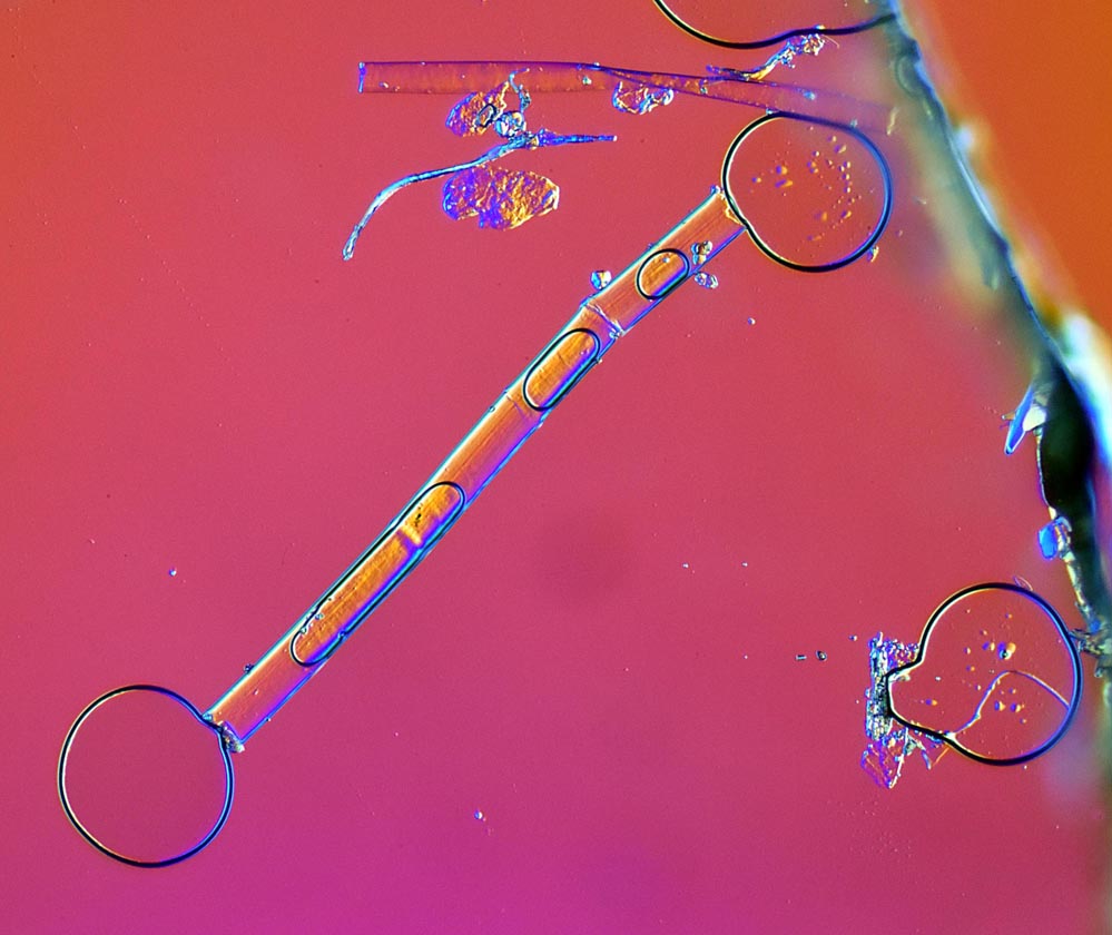
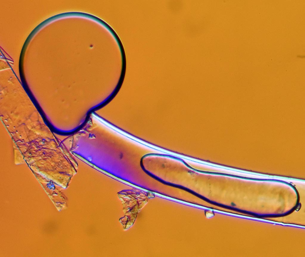
Finally, we get to transmitted DIC, here done on a Diaphot. A waveplate (retarder) can be inserted in the path, which is sometimes called a "tint plate". These first two images are with the tint plate installed, showing air bubbles escaping from small tubes (possibly dead algae, or some other biological source, most likely).

