Hoffman modulation contrast is similar to oblique illumination, in that the specimen is illuminated from one side of the pupil, as shown in the above figure (from the Olympus Microscopy Resource Center).
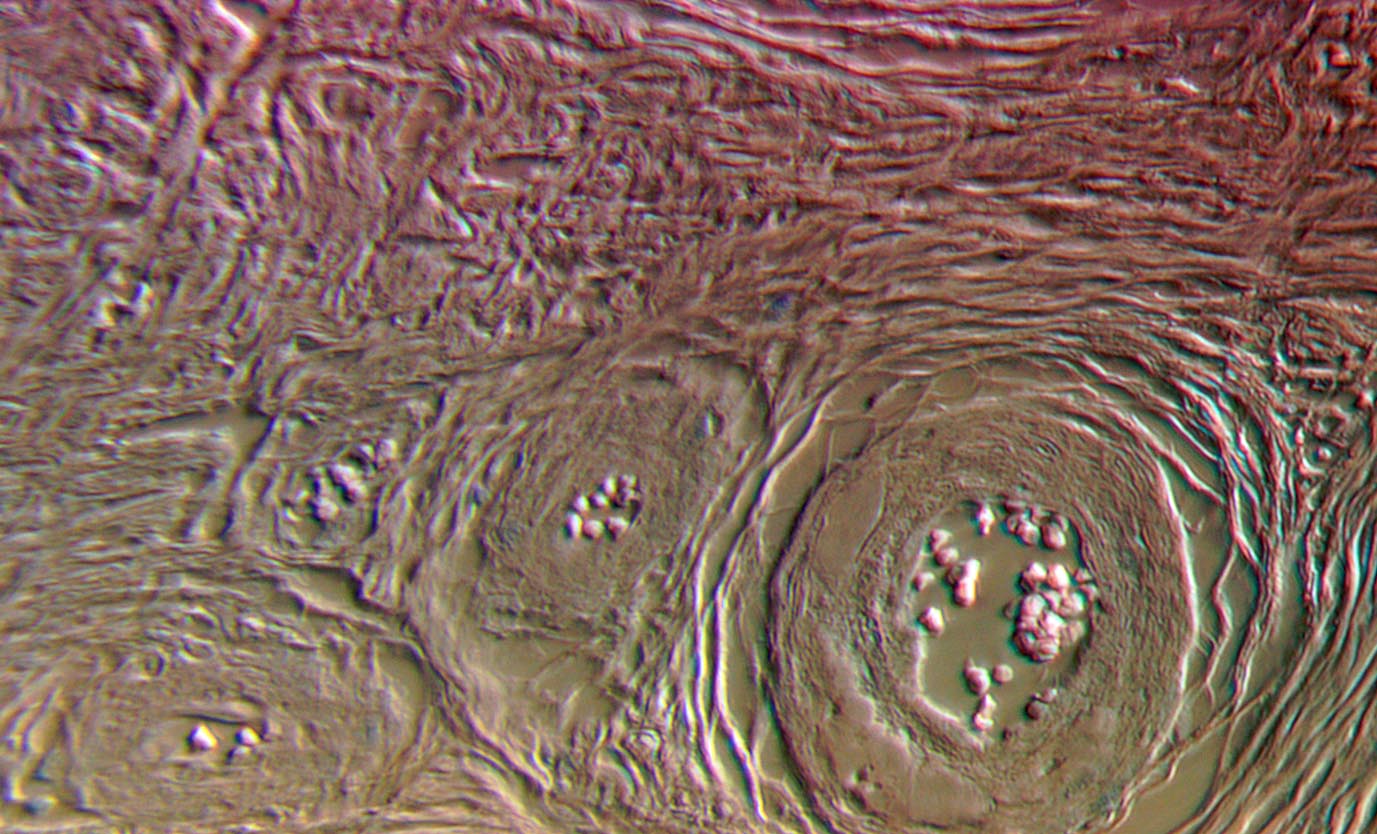
HMC images have a much different appearance than phase contrast, more like DIC or oblique illumination, which makes some sense since all three techniques have an "axis" of higher contrast. In the 20x images of a thin section of human cervical tissue below, Hoffman is represented on the left and phase contrast (Ph2 DL) on the right.


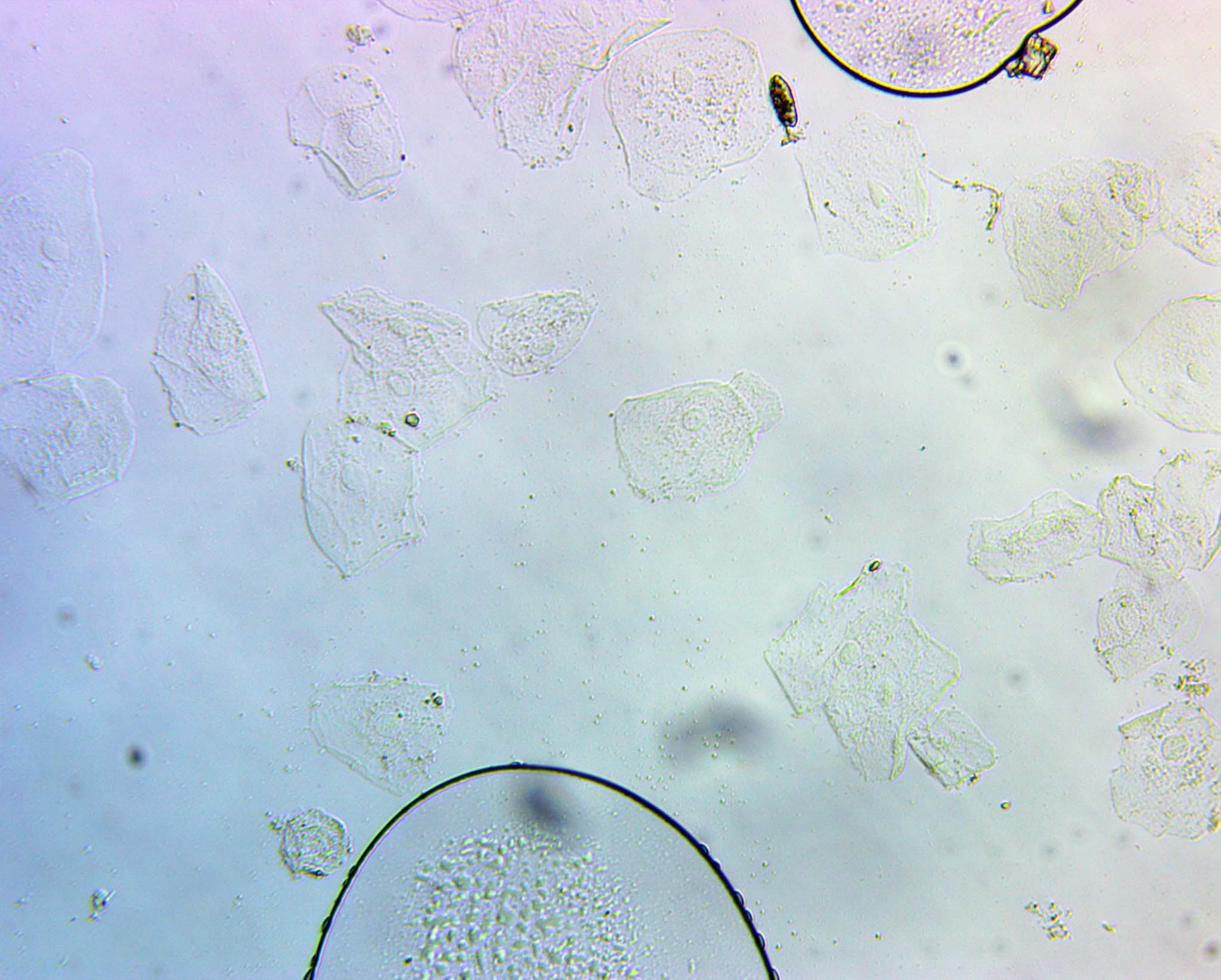
The above images are from a prepared slide, but if you are wondering where to get some fresh cells for observation, you fortunately have a vast supply - just grab a little rubbing from inside your cheek! Epithelial cells come off all the time in vast numbers, so are easy and painless to harvest. I use them here to demonstrate the power of Hoffman modulation contrast imaging with a simple Plan 10x.


First, to show how helpful this technique is, I took simple brightfield images, with the aperture open and then with it stopped down, reducing the NA into the system. The objective has an inherent (maximum) NA of 0.25. I then inserted the aligned (as shown above) HMC mask. The details pop right out, without the loss of resolution from reducing the NA! While each image could have been better with post-processing, I show it here only cropped.


I next went to the 20x objective (long working distance, NA = 0.40). In this case, instead of cropping, it is full-frame, but reduced in size by 50%. "Auto levels" were applied in this case in Photoshop.


And here is another region of the slide with the 20x HMC objective. This area is jam packed with cells.

In this next image, the axis of oblique illumination is the most readily apparent at the edge of the air bubble. It is bright in certain areas and dark in others - essentially like shadows from a setting sun illuminating a canyon on one side.
Overall, HMC is a simpler, cheaper method of getting good contrast on transparent specimens than DIC. It is a viable alternative, and has the benefit of being able to look through birefringent plates (like plastic petri dishes) without the odd effects DIC gives under these conditions.
The contrast can be adjusted by placing a polarizer between the light source and the condenser - on the same side of the specimen (with polarization and DIC observation, the specimen is between polarizers). This changes the intensity through the middle part of the HMC mask, adjusting image contrast.
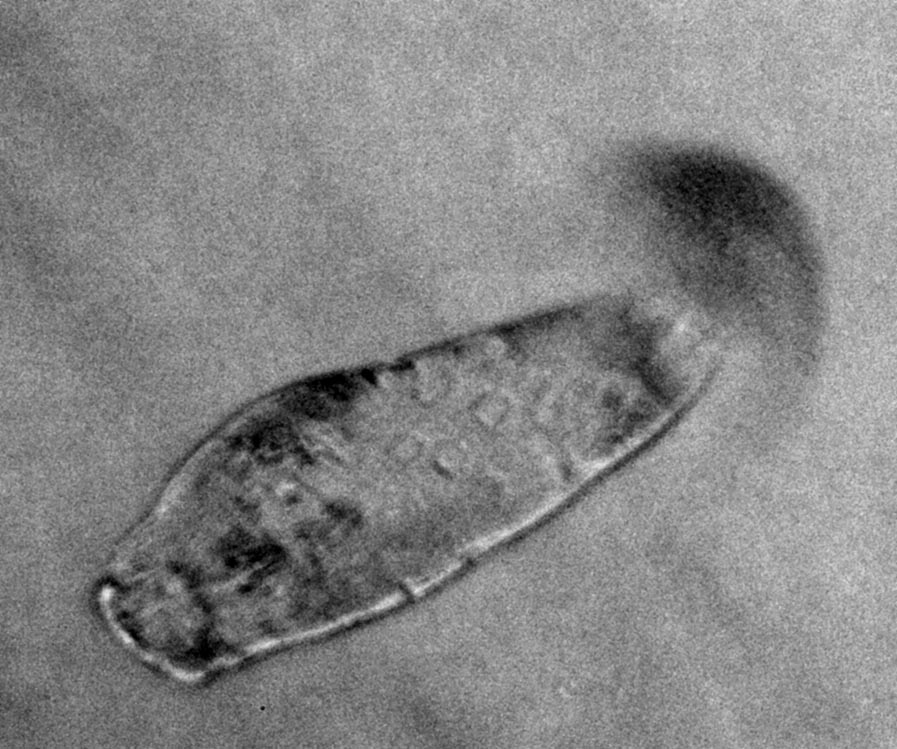
Here we see the death throes of an unfotunate protist, squished by surface tension by a cover glass.
 (13.5 MB .GIF)