 (5.5 MB .jpg)
Diatoms are one of the most interesting subjects. There are many varieties, and the details in each one is spectacular.
They often show up best in darkfield, phase contrast, and most of the other contrast enhancement techniques.
The large versions are availble by clicking on each thumbnail.
First we see a Victorian mount by C. Baker, London. Shown are three of the many in the strew in brightfield, and low-NA brightfield, darkfield and phase contrast, mostly at 40x.
The following are wider views of these diatoms, first at 10x and then 20x. They are: brightfield with very low NA, low NA, then darkfield and finally phase contrast (Ph1 and Ph2).
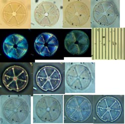
Next we have a Carolina Biological diatom test slide. Here we are at 100x phase contrast, comparing the phase ring densities of DL, DLL and DM (dark low, dark low-low, and dark medium).

Next is another Carolina Biological diatom, here in phase contrast at 40x Ph3 DL and 100x Ph4 DLL.


Here we see the Carolina Biological slide at 10x and 40x, brightfield and darkfield. These were taken with an Eclipse 200 and a homemade darkfield mask (a nickel, I think it was...). I think spherical aberration is seen, especially with the 40x, because I was trying a trinocular head for a Labophot/Optiphot, which doesn't contain the tube lens. It fits, and you can get an image, but the parfocality of the objectives is lost, and the images suffer.


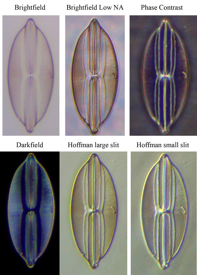
Now, I'll show the whole slide again at 20x (a single manual stitch required), in brightfield, BF with low NA, phase contrast, darkfield, and with two different slit widths of Hoffman modulation contrast (the first has the wider opening). This is on the finite conjugate Diaphot, rather than the Eclipse.
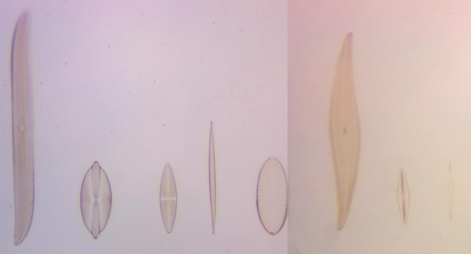
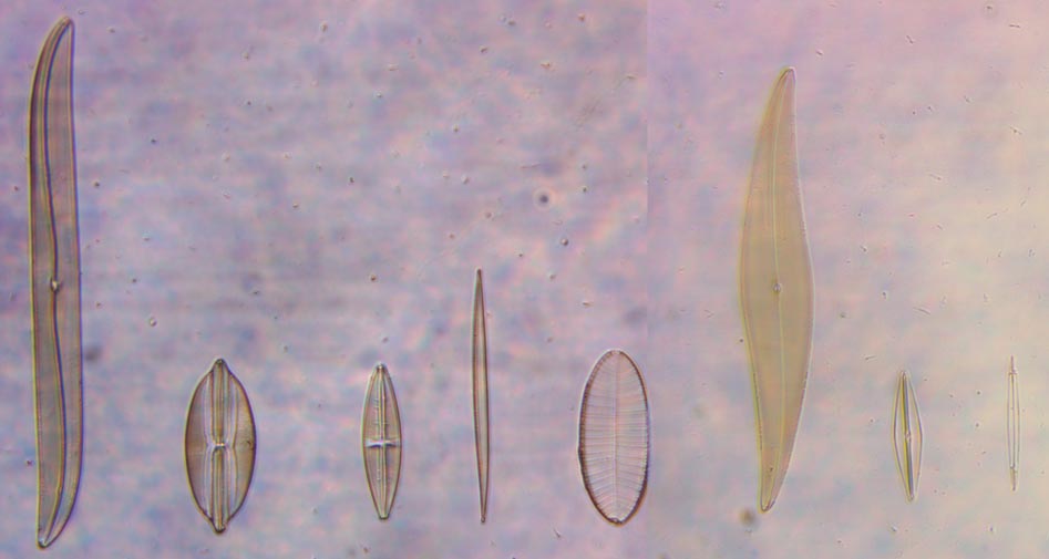
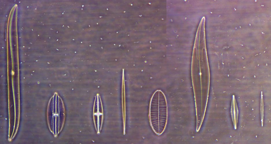
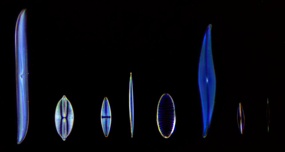
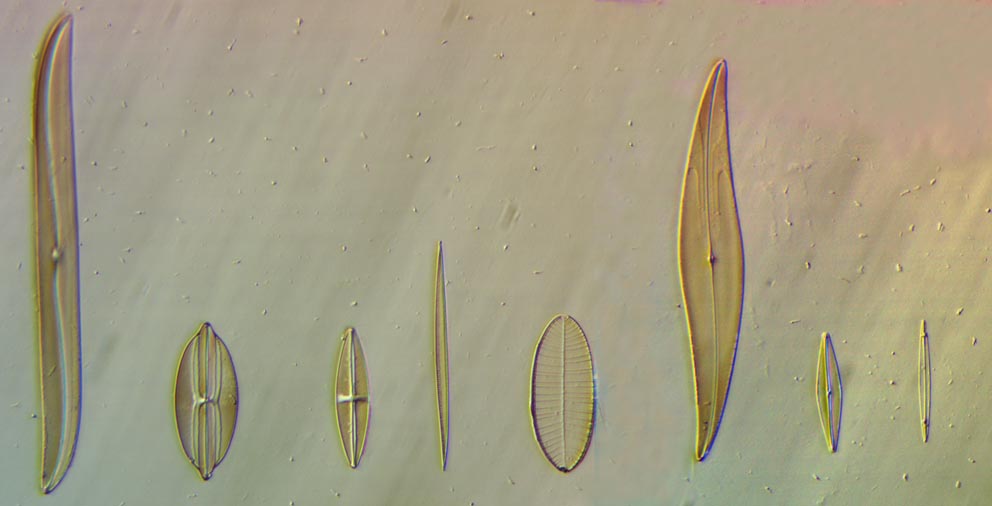
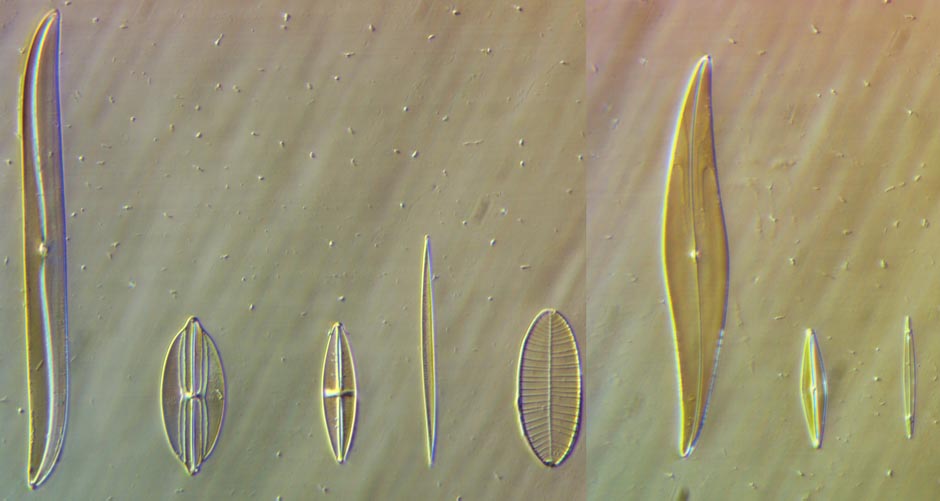
The above images are focus stacks of 2-5 images. Below are single images of one of the diatoms (#2 from the left).
