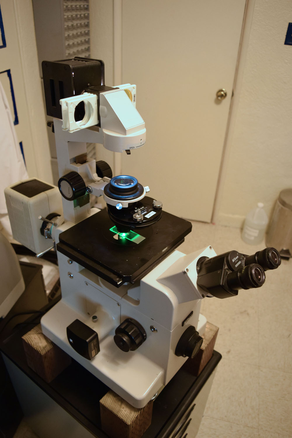
Here we see a Nikon Diaphot TMD with the Epi-Fluorescence (EF) attachment. The top light has the NCB blue filter and the excitation light from below is green. The images below were taken with an Optiphot 2 with EF and diascopic illumination.

The above photo is of a wing tip of a crane fly (mosquito eater), with crossed polarizers and darkfield providing the "background" and ultraviolet excited fluorescence making the blue/white highlights. This is at 10x with the UV-1A fluorescence cube installed.
Epi-fluorescence provides excitation light using the objective as a condenser as well (episcopic), with the object fluorescing at a longer wavelength. A dichroic beamsplitter passes the fluoresced light, but not the directly reflected excitation light. Furthermore, a barrier filter provides further extinction of the excitation bandwidth, providing the best contrast (signal-to-noise ratio). Capturing as much of the fluoresced light as possible, which is emitted in all directions, is essential to a good signal. To this end, very high NA objectives (for their respective power) are typically employed. For example, a typical 10x objective has an NA in the region of 0.25, but a Fluor objective is at a whopping 0.50!
\
Here is a ventral view of a tiny spider with a 10x objective, with green diascopic background light and bluish fluorescence, excited by UV light. By excited, I really mean that! The spider moved almost instantly when the UV lamp was unblocked. I think the small blue spots are blood cells, as I could see them moving around his body. I observed them going down to his feet quickly through the center of the legs and then returning more slowly around the edges.
I got some video of him reacting to the UV light (9.7 MB .wmv), as well as a longer one (134 MB .wmv) where he was staying put without the UV, but then moved. He didn't mind blue excitation as much, but eventually did move. His mouth is clearly visible, busy at first. Apparent blood flow is visible in both videos if you look closely.
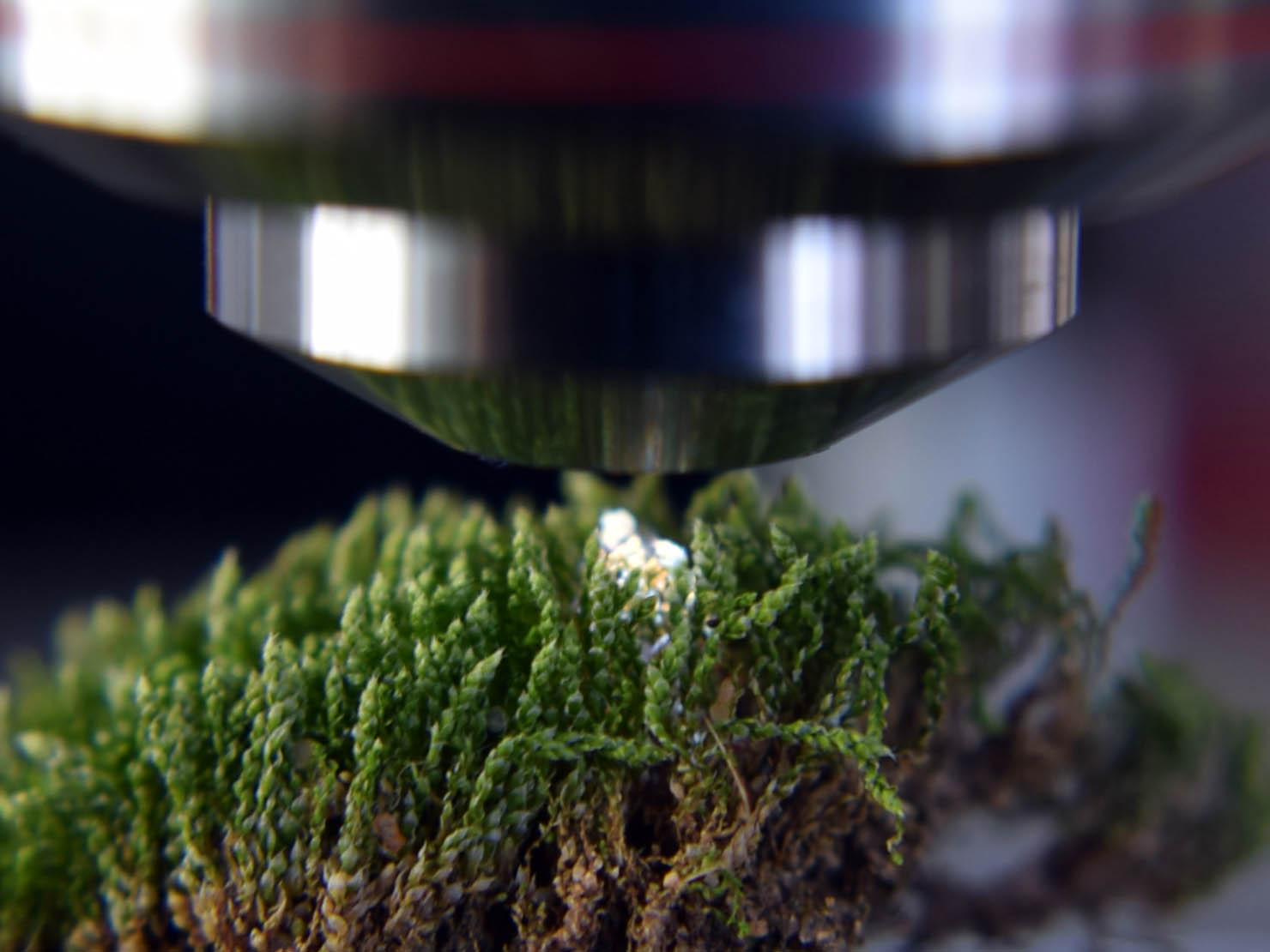
Fresh moss fluoresces brightly in UV light. The little leaves fluoresce white/blue and the buds at the end fluoresce deep red, but quickly (within ~20 seconds) photo-bleach and then fluoresce orange/yellow. Below is an external view under the scope. A tiny bit of orange is visible.
